Properties (11)
- purpleRed
The Motor Basis for Misophonia
Status:: #status/reviewed
Metadata
- Item Type: #journalArticle
- Authors: Sukhbinder Kumar, Pradeep Dheerendra, Mercede Erfanian, Ester Benzaquén, William Sedley, Phillip E. Gander, Meher Lad, Doris E. Bamiou, Timothy D. Griffiths
- Date Added: September 18, 2021
- URL: https://www.jneurosci.org/lookup/doi/10.1523/JNEUROSCI.0261-21.2021
- DOI: 10.1523/JNEUROSCI.0261-21.2021
- Cite Key: kumarMotorBasisMisophonia2021
- Tags: #Misophonia, #MRI, #Fmri
- PDF Attachments
Article Review table
| Background/Purpose | Background- • Misophonia is traditionally conceptualized as an emotional/auditory processing disorder • there exist neuron types that responds to the actions of others, the so called “Mirror neurons” • most triggers are related to orofacial movements/sounds • triggers can cause automictic mimicry in Misophonics • Misophonia involves distress to innocuous human-generated sounds. • Prior work showed anterior insula hyperactivity to trigger sounds. • Mechanism underlying why eating/chewing sounds evoke distress remains unclear. • Most triggers involve orofacial actions. • Mirror neuron system responds to observed and heard actions. • Spontaneous mimicry and emotional responses linked to MNS. Purpose- • test the authors hypothesis that the mirror neuron system related to orofacial movements plays a critical role in misophonia • specifically, test idea that the processes of mirroring triggers plays key role in miso • Test whether misophonia reflects “hyper-mirroring” of orofacial actions. • Examine resting-state connectivity between sensory and motor regions. • Examine sound-evoked connectivity and activation of orofacial motor cortex. • Determine relationship between motor activation and distress ratings. |
| Participants/samples | • Resting-state: N=33 (16 misophonia, 17 control after exclusions); age- and sex-matched. • Sound-evoked fMRI: N=42 (20 misophonia, 22 control); age- and sex-matched. • Misophonia inclusion: eating/breathing/chewing triggers; sound-alone sufficient; moderate–severe on Amsterdam scale. |
| Measures | • analysis of resting state fMRI connectivity and and sound evoked fMRI responses in Misophonics vs controls • Misophonia Questionnaire (Wu et al., 2014). • Amsterdam Misophonia Questionnaire. • Annoyance ratings (all subjects). • Misophonic distress ratings (misophonia group). • fMRI BOLD responses. • Resting-state functional connectivity. • Sound-evoked functional connectivity. |
| Procedures | • Resting-state: 10-minute eyes-open scan. • Sound-evoked: 3 sound categories (trigger, unpleasant, neutral). • 126 trials; 15 s sounds; post-sound ratings. • Seed-to-voxel connectivity analyses. • ROI: auditory cortex (HG, PT), visual cortex (V1, V2), vPMC, dPMC, anterior insula. • Group comparisons with age and sex covariates. |
| Results | • compared to controls, Misophonics were found to have no difference in response of auditory cortex to triggers but did find that: (1. Misophonics ha stronger resting state fMRI connectivity between the auditory and visual cortex and areas responsible for orifical movements; 2) Misophonics had generally stronger functional connectivity between auditory cortex and the orofacial motor area during sound perception; 3) Misophonics had stronger activation of orofacial motor area in response to trigger sounds. Resting-state connectivity • Misophonia showed stronger connectivity between right PT and right vPMC. • Stronger connectivity between right V2 and right vPMC. • Stronger connectivity between right vPMC and anterior insula. • Stronger connectivity between left anterior insula and M1/S1 and cerebellum. • No group differences in HG connectivity. • No differences in dPMC connectivity. Sound-evoked functional connectivity • Misophonia showed stronger connectivity between orofacial motor cortex and auditory cortex across all sound categories. • No group × sound interaction in auditory cortex connectivity. Activation • Orofacial motor cortex showed group × sound interaction. • Misophonia showed greater activation to trigger sounds only. • No trigger-specific activation differences in auditory cortex. • Orofacial motor activation increased proportionally with distress rating; stronger slope in misophonia. |
| Discussion/Limits | Conclusion- • results suggest that resposnes are the result of hyper mirroring of orofacial actions of other people. that is to say, there is excessive engagement of the orifical motor cortex via the auditory/visual sensory input associated with those actions. • the negative reactions to triggers, then, can bee seen as the result of annoyance / anger at being “forced” to “mimic” those actions. [see notes for more details] • based on this conclusion, misophonia should be though of as a sound issue, rather an issue of excessive mirroring with sound as the primary medium for said mirroring. this would also explain visual triggers. • data supports that mirroring of action underlies the anterior insula--based network in misophonia that has previously been observed. • The authors conclude that misophonia is characterized by increased coupling between sensory cortex and orofacial motor cortex. • The findings indicate excessive engagement of orofacial motor cortex in response to trigger sounds. • The data support a “hyper-mirroring” model in which sounds serve as a medium for mirroring actions. • The authors conclude that misophonia is not primarily a disorder of auditory cortex hyperactivity. • The findings indicate that anterior insula involvement may reflect mirroring-linked distress. • The authors conclude that therapies should target movement representations rather than sounds. |
| Limitations:: | • True implication of the mirror neuron system requires the use of single neuron recording methods that cant be done in humans ( outside of unique unique surgical circumstances) . as such all invocations of the mirror neuron system in this study are indirect. • the ongoing debate of the contributions of comorbid disorders to the symptoms of misophonia leave some questions about the distinctness of misophonia ( this should be addressed with studies using a mix of clinical and non clinical samples). • the mirror neuron hypothesis cant explain the occurrence of non-human/inanimate object triggers. it could be hypothesized that such triggers are introduced in tandem with normal triggers, resulting in a classically conditioned trigger, but such a prediction needs testing. • Evidence for mirror neuron involvement is indirect; fMRI lacks single-neuron resolution. • No clinical control group to assess disorder specificity. • Model does not directly test structural connectivity. • Less common non-orofacial triggers not directly examined. |
| Future Directions:: | • Use single-neuron recordings in rare clinical contexts. • Include clinical comparison groups. • Examine structural connectivity. • Test associative learning approaches targeting MNS. • Empirically test mechanisms for non-typical triggers. |
| Relevance:: | • the source of the Kumar model • this reconceptualization of misophonia as an mirroring issue rather then an emotional/auditory issue has drastic implications for our understanding of misophonia, the underlying neurology/etiology, and has drastic implications for future treatment attempts. |
| Statistics:: | • Resting-state exclusions: 1 misophonia (physiology unavailable); 3 controls (movement). • Age difference NS (p=0.38); sex difference NS (p=0.86). • Misophonia Questionnaire: 42.6±10.7 vs 17.6±8.8; p<0.001. • Amsterdam scale: 15.5±3.4 (misophonia). • Right PT–vPMC: peak 60,12,24; t=4.62; cluster=283; p<0.001; q(FDR)<0.05. • Right V2–vPMC: peak 58,0,24; t=4.34; cluster=221; p<0.001; q(FDR)<0.05. • vPMC–anterior insula: peak 42,6,6; t=4.64; cluster=165; p<0.001; q(FDR)<0.05. • Anterior insula–M1/S1: peak 18,16,78; t=4.82; cluster=383; p<0.001; q(FDR)<0.05. • Orofacial motor group×sound interaction: p=0.002 (SVC). • Rating slope difference: t(40)=6.8; p<0.001. |
Notes
key Terms
-
Misophonia – disorder characterized by strong negative emotions (anger, anxiety, disgust) in response to specific everyday sounds, particularly eating, chewing, breathing.
-
Trigger sounds – sounds that evoke misophonic reaction.
-
Mirror neuron system (MNS) – network including ventral premotor cortex that responds during both action execution and observation.
-
Ventral premotor cortex (vPMC) – region containing orofacial motor representations.
-
Anterior insula – region previously shown to correlate with misophonic distress.
my questions
my comments
- this idea is fascinating, and if true would drastically reshape not only how we conceptualize Misophonia, but how we might treat it.
misc. notes
-
based on the the required connectivity between the sensory and motor regions for the auditory/visuals/motor integration of mirror neurons ( described here ) guided the authors original goal of assessing the connectivity of the auditory and visual cortex to the rest of the brain in miso/control subjects
-
note on the order of of orifical motor cortex vs auditory cortex activation- looking at the trends in figure 3 Aii vs 3.B, shows that the increased connectivity between audiovisual sensory and orofacial motor areas in Misophonics is driven by greater misophonic reactivity in Misophonics to triggers, but not to unpleasant sounds. this specific increase for trigger sounds was not found for the auditory cortex, suggesting that activity starts in the orifical motor cortex, instead of starting with hyperactivity in the auditory system.
major Takeaways from paper
- supports idea that instead of miso being a sound/emotion disorder, it is an issue of hyper mirroring with sound acting as a vector.
Mirror neurons
- basics of Mirror neurons- a set of neurons that fire in response to the subject witnessing an action in anther individual. for example, there are mirror neurons in the official motor area that fire when we see someone move there mouth.
- defining feature of mirror neuron system ( auditory/visual/motor integration- in humans, this system associates auditory./visual patterns with apart of the motor cortex (ex: orifical motor cortex) involved in producing the input movement ( orofacial movements)
6 major characteristics of misophonia
(1) increased resting state functional connectivity
between the orofacial motor area and both auditory and visual
cortex; (2) increased functional connectivity between auditory
cortex and orofacial motor areas in response to all types of
sound; (3) increased activation of orofacial motor area in
response to trigger sounds specifically; (4) activation of orofacial
motor area increases in proportion to the misophonic distress;
(5) no difference from the control group in the activation of auditory cortex to trigger and other sounds; and (6) increased resting state functional connectivity between vPMC containing
orofacial motor area and insula in the resting state.“”
Authors major characteristics for Misophonia
future directions
- future studies should look into the possibility that Misophonics have stronger connectivity between the auditory and orofacial motor cortices .
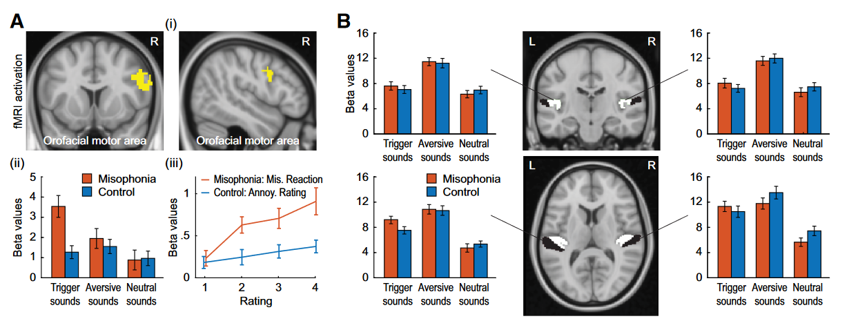
Figure 3. Activation of orofacial motor cortex and auditory cortex in response to three categories of sounds. Ai, The orofacial motor area represents a statistically significant (p =0.002) group sound category interaction. Aii, Bar plots represent b values for the orofacial motor cortex for the two groups in response to three sound categories. Aiii, Plots of variation of b values with the rating of misophonic distress in misophonia sufferers and of annoyance in control subjects. No group sound category interaction is seen in the auditory cortex (B,middlecolumn), which is confirmed by the bar plots of activation (B, first and third column) in response to sounds for the two groups. B, Middle column: white represents HG; black represents PT. Data in the bar plots represent mean 6 SEM.