Properties (15)
- purpleRed
- (Eijsker et al., 2021)
•
White matter abnormalities in misophonia
Status:: #status/Bare-bones-review
Metadata
- Item Type: #journalArticle
- Authors:: Nadine Eijsker, Arjan Schröder, Luka C. Liebrand, Dirk J.A. Smit, Guido van Wingen, Damiaan Denys,
- Date Added: August 25, 2021
- URL: https://linkinghub.elsevier.com/retrieve/pii/S221315822100231X
- DOI: 10.1016/j.nicl.2021.102787
- Cite Key: eijskerWhiteMatterAbnormalities2021
- Tags:: #Misophonia, #Bare-bones-review-🦴, #Neurology, #Fmri, #Attention, #White-matter-volume, #diffusion-tensor-imaging, #probablistic-tractography,
- PDF Attachments
Abstract
| Background/Purpose | Background- • Misophonia produces involuntary anger, anxiety, and/or disgust plus physiological arousal in response to specific ordinary sounds; prevalence estimates range from 3.2–6% clinically. • Low-intensity stimuli are proposed to become conditioned triggers via experience- and context-dependent processes. • Prior neuroimaging identified salience and emotional processing regions (anterior insula, cingulate, vmPFC, OFC, amygdala, hippocampus) as implicated during symptom provocation. • Attentional and behavioral abnormalities (Stroop impairment, aberrant alerting attention, response bias on stop signal task) exist outside symptom-provoking contexts. • Pre-attentive auditory processing is affected, evidenced by aberrant N1 ERP to non-trigger oddball sounds. • Only two prior structural studies exist: greater right amygdala GM volume (Eijsker et al., 2021) and greater vmPFC myelination (Kumar et al., 2017); no prior WM-focused structural analyses existed. • WM integrity is associated with global cognition and executive/motor functions; psychiatric disorders broadly show WM abnormalities. • Emotional disorders show reduced WM volumes and integrity; attentional/affective disorders show decreased WM integrity in attention-related tracts; ASD/sensory processing disorders show decreased WM integrity linked to abnormal auditory and multisensory processing. Purpose- • Overall goal: test for both macrostructural and microstructural WM differences between misophonia patients and healthy controls using T1-weighted MRI and DTI. • Secondary goal: determine whether WM structure correlated differently with age between groups. • Tertiary goal: examine whether WM structural abnormalities had linear relationships with clinical symptom characteristics in patients. |
| Participants/samples | • 24 misophonia patients; 25 matched controls. • Adults (18–65); matched on age, sex, education. • Exclusions: major psychiatric disorders, neurological conditions, substance disorders, hearing loss. • Patients diagnosed per Schröder et al. (2013) criteria by three experienced AMC psychiatrists. • Inclusion criterion for patients: anger (and in some cases disgust) in reaction to eating sounds and at least three of four specified sounds. • Exclusion criteria (all participants): major depression, anxiety disorder, bipolar disorder, psychotic disorder, ASD, substance-related disorder, hearing loss, epilepsy, structural CNS disorder, stroke within prior year, MRI contraindications. |
| Measures | • A-MISO-S: misophonia symptom severity (adapted Y-BOCS). • SCL-90: general psychopathology. • HAM-A: anxiety severity. • HAM-D: depression severity. • BPAQ (Buss-Perry Aggression Questionnaire): physical aggression, verbal aggression, anger, hate, and total aggression subscales. • SCID-II: personality disorder screening. • DTI measures: FA, MD, RD, AD (averaged within FA skeleton and voxel-wise via TBSS). • VBM: WM volume maps from T1-weighted images. |
| Procedures | • Whole-brain VBM to assess WM volume differences. • DTI preprocessing and tract-based spatial statistics (TBSS) for voxel-wise analysis. • Probabilistic tractography seeded from VBM peak voxels. • Correlation analyses with symptom severity and clinical measures. |
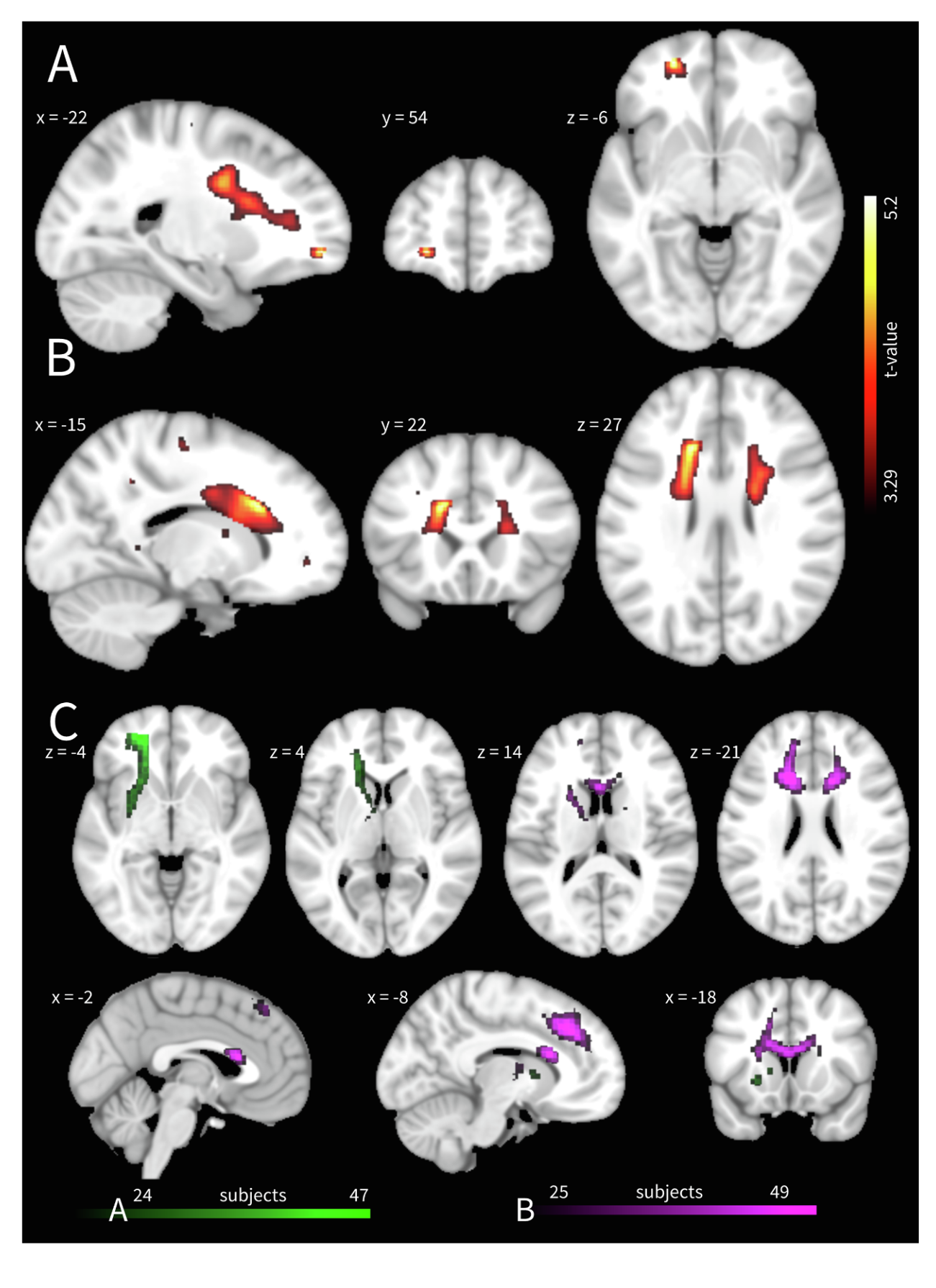
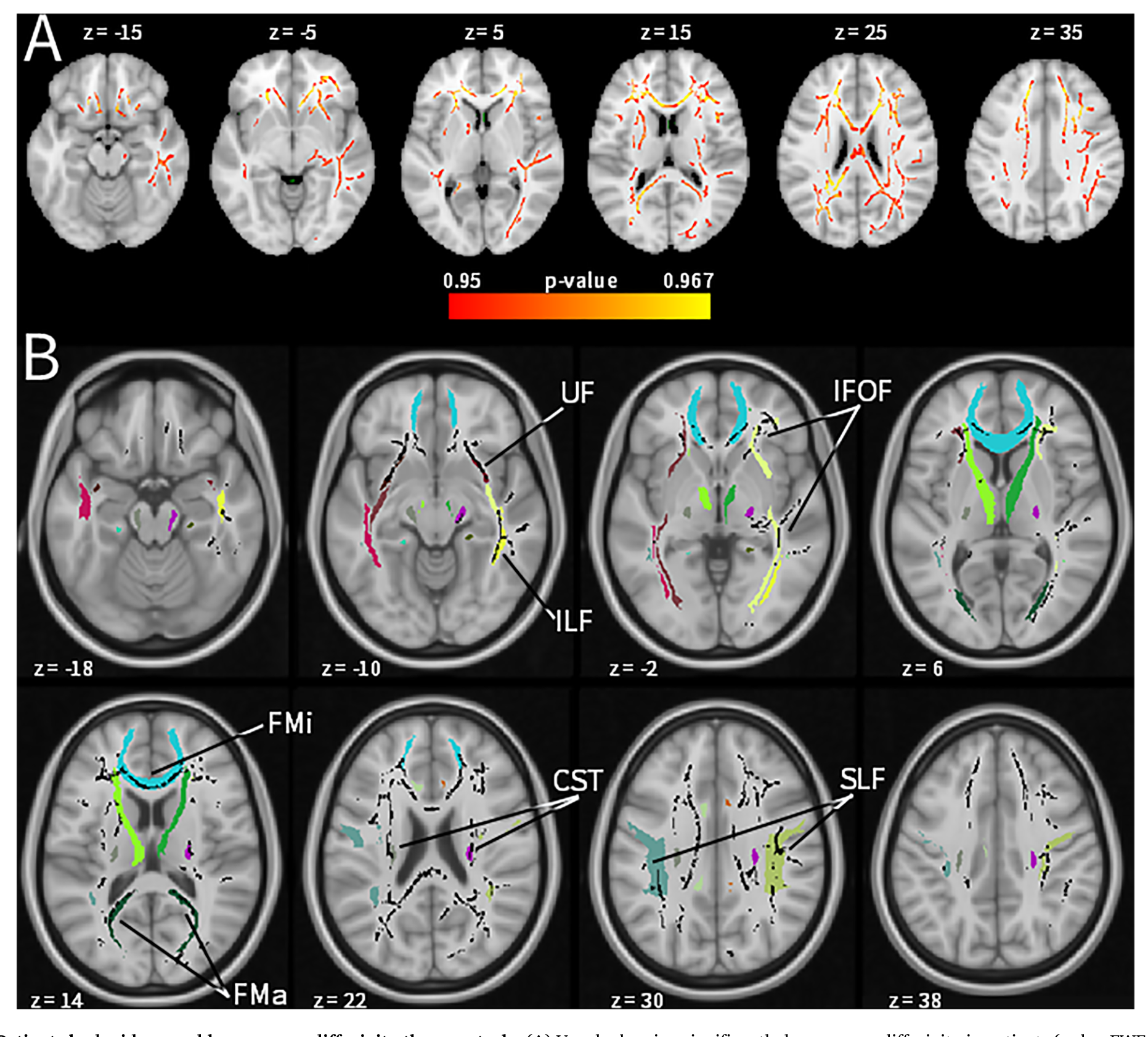
| Results | Macrostructural WM (VBM) • Patients had greater left-hemispheric WM volumes at two frontal locations compared to controls. • Probabilistic tractography from the anterior peak voxel implicated the left IFOF and ventromedial ATR. • Tractography from the body of the corpus callosum (BCC) peak implicated bilateral connections to superior frontal gyri and the dorsolateral ATR. • No group × age interactions for WM volume. Microstructural WM (DTI) • Patients had lower averaged MD and RD within the FA skeleton compared to controls; medium-to-large effect sizes. • Averaged FA showed a medium-to-large but non-significant trend toward higher values in patients; averaged AD showed a non-significant trend. • Voxel-wise TBSS indicated bilateral, widespread lower MD in patients spanning the IFOF, uncinate fasciculus (UF), ATR, inferior and superior longitudinal fasciculi (ILF, SLF), and forceps minor and major. • No group × age interactions for DTI measures. • Exclusion of the three scans acquired with fewer diffusion directions did not change the pattern of significant findings. Correlational analysis • WM volume at either peak voxel (IFOF/ATR or BCC/ATR) and mean MD showed no significant linear relationship with A-MISO-S, HAM-A, or BPAQ anger after correction. |
| Discussion/Limits | Conclusion- • The authors conclude that misophonia is associated with both macrostructural (greater left frontal WM volume) and microstructural (lower MD and RD) white matter abnormalities, supporting a neurobiological basis. • The findings indicate that reduced MD and RD likely reflect increased axonal density and myelination, consistent with higher WM integrity. • The authors conclude that affected tracts (IFOF, UF, ATR, ILF, SLF, BCC) link frontal, parietal, occipital, and subcortical regions and subserve social-emotional processing and attentional orienting. • The findings indicate that abnormalities in social-emotional processing, particularly facial emotion recognition, align with the social nature of trigger sounds and prior amygdala and fusiform findings. • The authors conclude that WM alterations in attention-related networks correspond to heightened orienting toward aversive stimuli and difficulty disengaging attention. • The data support that altered white matter, including in forceps minor, may relate to sensory enhancement of trigger-related input via attention networks. • The authors conclude that these alterations may underlie the neuropsychological profile of misophonia, though further research is required to establish its classification as a discrete disord |
| Limitations:: | • Small sample size limits generalizability and statistical power; effects with considerable effect sizes did not reach significance. • Whole-brain exploratory analysis with rigorous multiple comparison correction may have missed effects detectable with region-of-interest approaches (e.g., vmPFC alterations found by Kumar et al. 2017). • Some participants scanned with fewer diffusion directions (48 vs. 64), though sensitivity analysis confirmed this did not affect results. • No reverse-phase encoded scans acquired, precluding susceptibility-derived distortion correction. • Two comorbid ADHD patients and one with borderline personality disorder included; clinical heterogeneity not fully controlled. |
| Future Directions:: | • Replication of current results in larger samples is warranted. • Future studies should relate WM alterations to differences in emotional reactivity, attentional functioning, and cognitive control, including the effect of symptom provocation. • Direct investigation of trigger-related visual scene processing is proposed, with primary focus on human faces given the prevalence of nasal and oral misophonia triggers. |
| Relevance:: | • study looked at structural abnormalities in white matter in Misophonia, finding abnormalities that they conclude maybe indicative of Misophonia • an example of fMRI analysis applied to Misophonia. |
Notes
key terms and concepts
-
Voxel-based morphometry (VBM) – A brain scanning technique that compares the size and volume of specific tiny regions of brain tissue between groups of people. Think of it like comparing the thickness of different sections of a map grid square by square.
-
Diffusion tensor imaging (DTI) – A type of MRI that tracks how water molecules move through the brain's white matter "cables." Healthy, well-organized cables restrict water movement in predictable ways, so unusual water movement patterns signal something is different about the tissue.
-
Fractional anisotropy (FA) – A measure of how directionally organized the water movement is inside a brain cable. High FA means water flows in one tidy direction (like a well-bundled wire), suggesting healthy, intact tissue.
-
Mean diffusivity (MD) – The overall average speed at which water spreads in all directions. Lower MD generally means the tissue is more tightly packed and dense.
-
Radial diffusivity (RD) – How much water spreads sideways across a brain cable (perpendicular to its length). Lower RD tends to suggest thicker insulation (myelin) around the nerve fibers.
-
Axial diffusivity (AD) – How much water travels lengthwise along a brain cable. Changes here can reflect damage to the cable itself.
-
Tract-based spatial statistics (TBSS) – A standardized method for comparing DTI measurements across participants by projecting everyone's data onto a shared "skeleton" map of the brain's major white matter pathways, then testing every point on that map for group differences.
-
Probabilistic tractography – A computational method that traces likely pathways of brain "wiring" by following the most probable routes water would travel. It's essentially a GPS for mapping which brain regions are connected to each other.
-
Inferior fronto-occipital fasciculus (IFOF) – A long white matter bundle running from the front of the brain (involved in decision-making and emotion) all the way back to the visual processing areas at the rear. It plays a role in recognizing facial expressions and emotions.
-
Anterior thalamic radiation (ATR) – A set of connections between the front of the brain (prefrontal and orbitofrontal cortex) and the thalamus, a central relay hub. This pathway helps regulate automatic attention to emotional stimuli and voluntary focus on relevant tasks.
my questions
my comments
Bookmarks
reading notes
contextual statement
In a 2021 study, Eijsker attempted to characterize macro- and microstructural white matter abnormalities in misophonia by comparing 24 patients and 25 matched healthy controls using T1-weighted VBM, DTI with TBSS, and probabilistic tractography.
zettlekasten
findings of white matter structural abnormalities in Misophonia